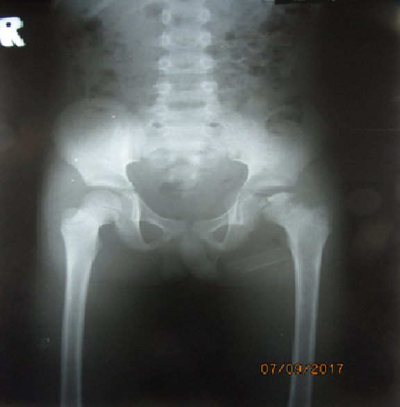
History: An 11 year old boy came to CSC with his mother. His mother describes the boy as having left hip pain for two months, with no history of trauma. The pain did not improve with pain killers and they first went to Kuntheabopha hospital. An x-ray was taken and the patient referred to CSC. The child does not have any other medical problems in the past.
The child however says he does not have any pain in the hip when he arrives to CSC. He denies any history of fever.
Dr Saqib: Learning point 1:There are many causes of hip pain in children, and usually taking a good history from the patient will give most of the answers. Causes of hip pain include development dyplasia of the hip (DDH), septic arthritis, transient synovitis, Perthes disease, trauma, slipped upper femoral epiphysis (SUFE), metabolic disease and even rarely, tumour. Sometimes hip pain can be coming from other parts of the body like a problem in the spine or knee. Normally asking questions about the hip pain will help give the diagnosis, like at what age the hip pain started, how quickly the problem developed and whether it is associated with symptoms like fever or trauma. It is very important at CSC you take a full history about the hip pain before examining the child.
This Youtube video helps gives an overview of the different conditions but is very basic. Please also read
these guidelines.
When examining a child, it is important to do a full examination. When we examined this patient, he walked normally with no deformity, no leg length discrepancy, and no areas of tenderness. However, when we assessed the range of motion, when the child was lying down, there was restriction of movement when the hip was flexed and internally rotated.
We then ordered an x-ray of the patient’s hip. It is good to get an AP Pelvis and a lateral / frog lateral of the affected hip.

Dr Saqib: Learning point 3: Here are on this x-ray we can see flattening of the femoral head on the left hand side, with some sclerosis and subchondral lucency. With the history, examination and x-ray, we can now diagnose this as Perthes disease. Please read
this link to learn more about Perthes disease. Dr Saqib: Learning point 4: Perthes disease goes through a number of stages, starting from Initial, Fragmentation, Reossification and Remodelling. This is the Waldenstrom Stages. The objective of the treatment is to allow the femoral head the best chance of re-ossification and remodelling in the correct position.
Watch this full overview which gives advance discussion on management.
Or you can read the article below, Management of Perthes’ disease – Benjamin Joseph:
[gview file=”http://teaching.csc.org/wp-content/uploads/2017/09/joseph2015.pdf”]
